TMS and Brain Mapping
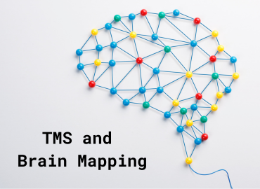
The cells of the brain are known as neurons. The brain is constantly humming with communication between these neurons. They receive and send messages to each other and all the other parts of our body in electrical impulses we call “brain waves.” These can be made visible with a procedure called an electroencephalogram, or EEG. They provide a literal visual map of the brain.
The Society for Brain Mapping and Therapeutics (SBMT) defines brain mapping as, “The study of the anatomy and function of the brain and spinal cord through the use of imaging, immunohistochemistry, molecular and optogenetics, stem cell and cellular biology, engineering, neurophysiology, and nanotechnology.”
To map the brain, an EEG is administered using a sensory instrument placed on the patient’s scalp. Software connected to the device displays brain wave patterns on a screen. Brain waves are categorized into Delta, Theta, Alpha, and Beta waves. Doctors can then identify any dysfunctional areas in the lobes of the brain, which are the frontal, parietal, central, temporal, and occipital lobes.
A brain map is a fantastic tool for finding places in the brain that are not functioning correctly and understanding what is going on. Being able to map the brain has greatly advanced the speed and accuracy of data collection from brain waves. The goal is to improve communication between the areas of the brain and the rest of the body. The patient can also work with the doctor to self-assess their emotional and cognitive function so that it can be compared with the brain map’s findings.
We use the noninvasive brain stimulation treatment TMS (transcranial magnetic stimulation) at this clinic to help patients with major depressive disorder (MDD) lead healthier lives. Brain mapping lets us zero in on the left dorsolateral prefrontal cortex (DLPFC), located near the left temple. That is where we apply the coil through which flows the electromagnetic current that stimulates the mood centers of the brain.
The DLPFC also helps govern working memory, decision-making, and emotional response to stimuli. This portion of the brain is closely linked to the limbic system, which also governs emotional and behavioral responses to stimuli. People with depression tend to have dysfunctional and underactive activity in the left dorsolateral prefrontal cortex.
For more information relating to this topic, checking out these posts:
− TMS and electromagnetism in the human brain
− Characteristics of the depressed brain
For questions and appointments, write to us on our website or call (585) 442-6960.


